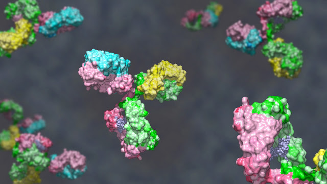
Anti-nuclear antibody testing is a popular test used to diagnose autoimmune disorders

Anti-nuclear Antibody Testing
Anti-nuclear antibody testing is
a blood test used to look for autoantibodies that attack components of cells’
nuclei, or "command" centers. These antibodies can trigger autoimmune
diseases like lupus. Some people who have autoimmune disorders such as lupus or
Sjogren's syndrome will have antibodies that specifically target the DNA in
their cells' nuclei. These autoantibodies are called ANAs.
According to Coherent Market Insights the Anti-nuclear
Antibody Testing Market Global Industry Insights, Trends, Outlook, and
Opportunity Analysis, 2022-2028.
There are several ways to measure
ANAs in a person's blood. The most common method is the indirect
immunofluorescent ANA (IIF-ANA) test, also known as a fluorescent antinuclear
antibody (FANA) test. Some of the supplemental tests that are commonly ordered
with a positive ANA include a Sjogren's syndrome test, an anti-double-stranded
DNA test (anti-dsDNA), and a drug-induced SLE (anti-U1RNP) test. These tests
help the doctor confirm a diagnosis of an autoimmune disorder and may rule out
other causes for symptoms, including infection, cancer, or drugs. There are
many different types of autoimmune disorders, each with its own set of signs
and symptoms. Symptoms can include joint pain, swelling, and fever. Some of
these diseases are more serious, like rheumatoid arthritis and scleroderma.
The ELISA test is the most
sensitive of the anti-nuclear antibody
testing and can detect single autoantibodies such as anti-dsDNA, SS-A/Ro,
and SS-B/La. The test is based on the principle that an antibody binds to its
epitope (target) in a solid phase well when it is stimulated by an enzyme. The
test uses a technique called indirect immunofluorescence assay, or IFA. This method
allows ANA in patient serum to bind to corresponding nuclear epitopes on
microscope slides of human epithelium-derived HEp-2 cells. This is followed by
the detection of the resulting fluorescent signal with an enzyme-conjugated
detection antibody. The intensity of the fluorescent staining and the pattern
of binding is then analyzed at a series of dilutions to determine a level of
ANA that is most likely to be present in the sample.


Comments
Post a Comment